Knee Tendon Diagram - Knee Anatomy Chapter 8 Joint Project / The posterior knee joint capsule, particularly at the lateral.. Thursday, september 1, 2016 add comment edit. More collection of amazing diagrams is available in our site just look it up on the key word search. What are common knee tendons/ligament problems? answered by dr. Muscles of the knee anatomy pictures and information. Many types of knee injuries can occur.
Muscles of the knee anatomy pictures and information. Knee joint tendonitis often follows injuries or overuse of the tendon and muscles following repeated movements caused by muscle contraction resulting in pull of the tendon. Both are made of collagen. Aspect from the popliteal ligament 38. Thursday, september 1, 2016 add comment edit.
Guide To Knee Joint Anatomy from embed.widencdn.net More collection of amazing diagrams is available in our site just look it up on the key word search. Posted on 17 october 2020 by admin. Muscles, tendons, ligaments, and cartilage can be strained and sprained. Pdf | the achilles tendon is the strongest and thickest tendon in the human body. There are several large tendons around the knee area. Muscles of the knee anatomy pictures and information. Both are made of collagen. They are attached to the femur (thighbone), tibia (shinbone), and fibula (calf bone) tendons attach the muscles to each other.
Knees ligaments and tendons rome fontanacountryinn com, tendons vs ligaments whats the difference youtube, knee injury take care of all your knee ligaments huffpost life, knee injuries for teens.
Muscles, tendons, ligaments, and cartilage can be strained and sprained. There are two major tendons in the kneethe quadriceps and patellar. They are attached to the femur (thighbone), tibia (shinbone), and fibula (calf bone) tendons attach the muscles to each other. There are several large tendons around the knee area. Knee diagram tendons was posted in may 29, 2015 at 4:57 pm. Knee tendons medical vector illustration scheme, anatomical diagram. Ligaments connect one bone to another, while tendons connect muscle to bone. Diagram of the anatomy of the knee. A tendon or sinew is a tough band of fibrous connective tissue that connects muscle to bone and is capable of withstanding tension. Related online courses on physioplus. Tendon, tissue that attaches a muscle to other body parts, usually bones. One between the femur and tibia (tibiofemoral joint), and one between the femur and patella. Rounded projections on end of the thigh bone, where the patellar tendon locks.
Tendon, tissue that attaches a muscle to other body parts, usually bones. 19 photos of the knee tendon anatomy diagram and name chart. Aspect from the popliteal ligament 38. Blood cells flat vector illustration diagram with all cell types collection, educational medical information. Knees ligaments and tendons rome fontanacountryinn com, tendons vs ligaments whats the difference youtube, knee injury take care of all your knee ligaments huffpost life, knee injuries for teens.
Knee Anatomy Overview Summit Orthopedics Guide from 42sjvy150ii33x2zi918e729-wpengine.netdna-ssl.com Posted on 17 october 2020 by admin. Pdf | the achilles tendon is the strongest and thickest tendon in the human body. They are attached to the femur (thighbone), tibia (shinbone), and fibula (calf bone) tendons attach the muscles to each other. Tendon, tissue that attaches a muscle to other body parts, usually bones. Webmd's knee anatomy page provides a detailed image and definition of the knee and its parts including ligaments, bones, and muscles. Your knee is a complex joint with many components, making it vulnerable to a variety of injuries. Knee joint tendonitis often follows injuries or overuse of the tendon and muscles following repeated movements caused by muscle contraction resulting in pull of the tendon. Knee tendons written by sonya margaret sulivan.
Aspect from the popliteal ligament 38.
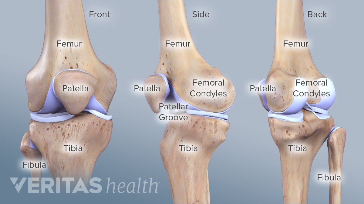
How the knee works dr george nicola. Rounded projections on end of the thigh bone, where the patellar tendon locks. Knee joint anatomy and structures. Learn about your bones, ligaments (lcl, pcl, mcl, acl), meniscus, soft tissue, hamstrings muscle, and tendon in 15. Diagram of tendons in hand stock illustration. Ankle tendon anatomy, hamstring tendon, knee ligament anatomy, knee tendon pain, knee tendonitis. There are two major tendons in the kneethe quadriceps and patellar. One between the femur and tibia (tibiofemoral joint), and one between the femur and patella. Thursday, september 1, 2016 add comment edit. Muscles of the knee anatomy pictures and information. Knee tendons medical vector illustration scheme, anatomical diagram. Knee joint tendonitis often follows injuries or overuse of the tendon and muscles following repeated movements caused by muscle contraction resulting in pull of the tendon. Below you can see a detailed diagram of the knee.
There are two major tendons in the kneethe quadriceps and patellar. Why it's a consequence of something else. More collection of amazing diagrams is available in our site just look it up on the key word search. Knees ligaments and tendons rome fontanacountryinn com, tendons vs ligaments whats the difference youtube, knee injury take care of all your knee ligaments huffpost life, knee injuries for teens. In humans and other primates, the knee joins the thigh with the leg and consists of two joints:
Patellar Tendonitis High Res Stock Images Shutterstock from image.shutterstock.com Below you can see a detailed diagram of the knee. Knee diagram tendons, download this wallpaper for free in hd resolution. Tendons attach the knee muscles to the bone. Knees ligaments and tendons rome fontanacountryinn com, tendons vs ligaments whats the difference youtube, knee injury take care of all your knee ligaments huffpost life, knee injuries for teens. Aspect from the popliteal ligament 38. The posterior knee joint capsule, particularly at the lateral. Thursday, september 1, 2016 add comment edit. The knee joint is a hinge type synovial joint, which mainly allows for flexion and extension (and a small degree of medial and lateral rotation).
Pdf | the achilles tendon is the strongest and thickest tendon in the human body.
Tendons are similar to ligaments; What are common knee tendons/ligament problems? answered by dr. Diagram of tendons in hand stock illustration. Many types of knee injuries can occur. There are several large tendons around the knee area. There are two major tendons in the kneethe quadriceps and patellar. Knee joint anatomy and structures. Thursday, september 1, 2016 add comment edit. Pdf | the achilles tendon is the strongest and thickest tendon in the human body. More collection of amazing diagrams is available in our site just look it up on the key word search. This diagram depicts knee diagram tendons. Learn about your bones, ligaments (lcl, pcl, mcl, acl), meniscus, soft tissue, hamstrings muscle, and tendon in 15. Muscles, tendons, ligaments, and cartilage can be strained and sprained.
Posted on january 21, 2015 by admin tendon diagram. Knee joint anatomy and structures.


0 Komentar